Finite element analysis of compression fractures at the
By A Mystery Man Writer
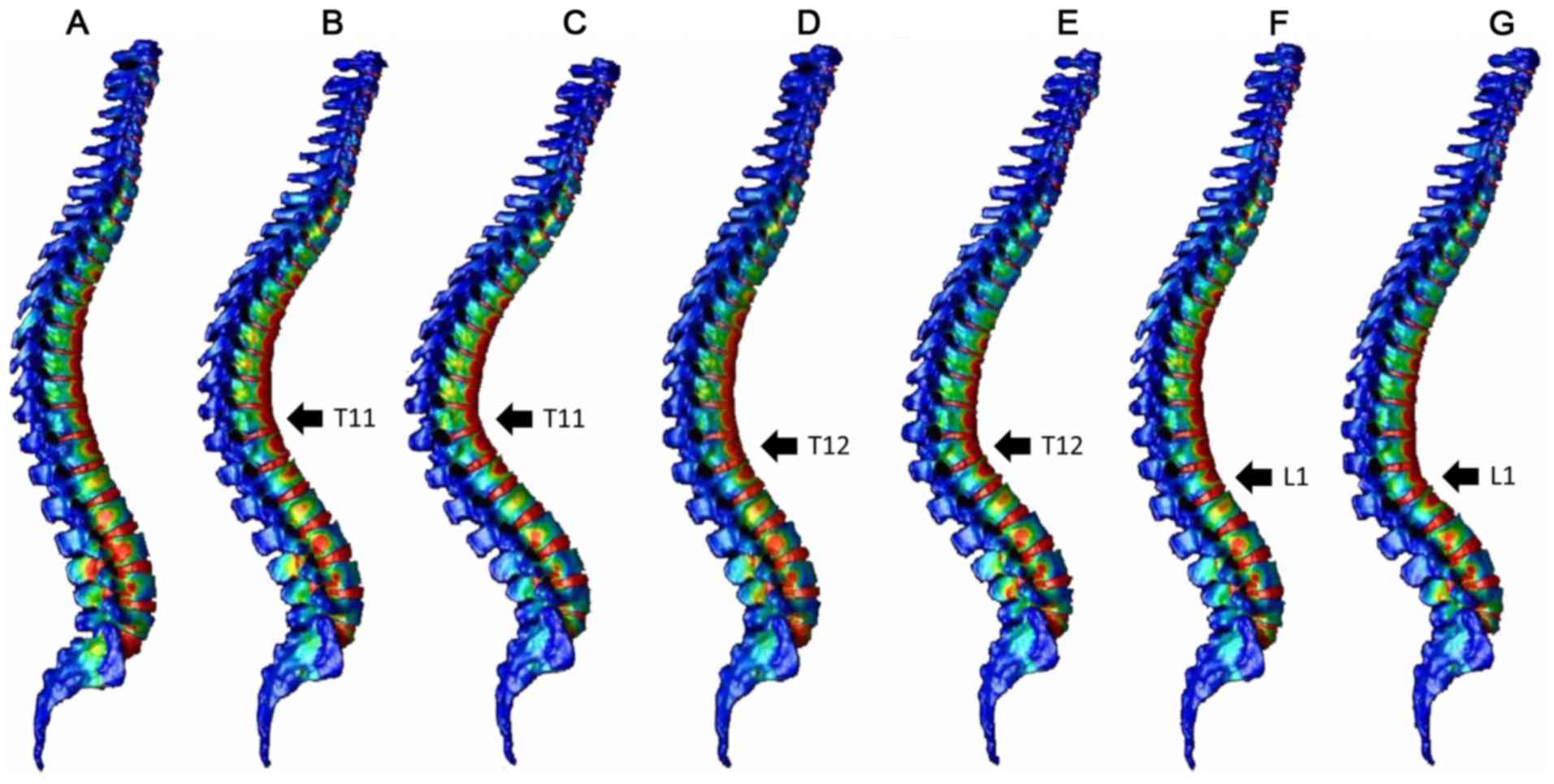
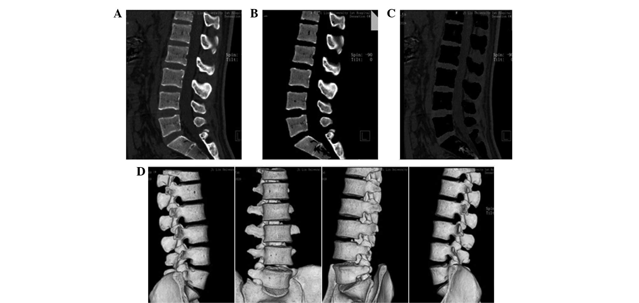
Vertebral fractures commonly occur at the thoracolumbar junction. These fractures can be treated with mild residual deformity in many cases, but are reportedly associated with increased risk of secondary vertebral fractures. In the present study, a three‑dimensional (3D) whole spine model was constructed using the finite element method to explore the mechanism of development of compression fractures. The 3D model of the whole spine, from the cervical spine to the pelvis, was constructed from computed tomography (CT) images of an adult male. Using a normal spine model and spine models with compression fractures at the T11, T12 or L1 vertebrae, the distribution of strain was analyzed in the vertebrae after load application. The normal spine model demonstrated greater strain around the thoracolumbar junction and the middle thoracic spine, while the compression fracture models indicated focused strain at the fracture site and adjacent vertebrae. Increased load time resulted in the extension of the strain region up to the middle thoracic spine. The present findings, that secondary vertebral fractures commonly occur around the fracture site, and may also affect the thoracic vertebrae, are consistent with previous clinical and experimental results. These results suggest that follow‑up examinations of compression fractures at the thoracolumbar junction should include the thoracic spine and adjacent vertebrae. The current data also demonstrate that models created from CT images can be used for various analyses.

Finite Element Analysis and Its Applications in Dentistry
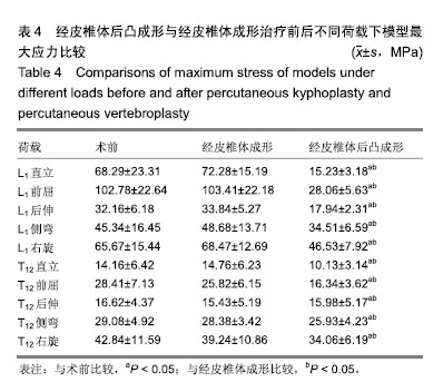
Mechanical changes of percutaneous kyphoplasty and percutaneous vertebroplasty in the treatment of thoracolumbar compressive fractures in three-dimensional vertebral models

PDF] Finite element analysis of compression fractures at the thoracolumbar junction using models constructed from medical images
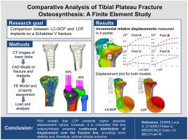
Comparative analysis of tibial plateau fracture osteosynthesis: A finite element study,Journal of the Mechanical Behavior of Biomedical Materials - X-MOL

Finite Element Analysis of Vertebral Compression Fracture
Novel, fast and efficient image‑based 3D modeling method and its application in fracture risk evaluation

Computer-Assisted Quantification

Finite element analysis of double‐plate fixation using reversed locking compression‐distal femoral plates for Vancouver B1 periprosthetic femoral fractures, BMC Musculoskeletal Disorders

Finite Element Analysis of Fracture Fixation
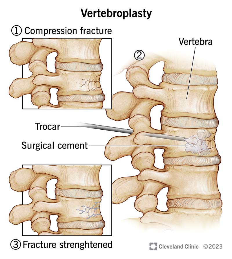
- Vertebroplasty: What It Is, Purpose, Procedure & Side Effects
- Management of vertebral compression fractures - Sports Medicine Review

- Compression Fracture Treatment, Causes, & Symptoms

- Frontiers Case report: Use of peripheral nerve stimulation for
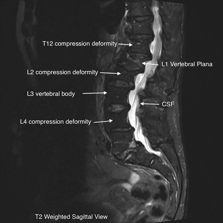
- An 80-year-old female patient with thoracic 12 vertebral compression

- The Denim jumpsuit- Light wash – marsthelabel

- How to Use a Compass [EASY Step-by-Step Tutorial] – Greenbelly Meals

- Hanes Women's Premium Boyfriend Ring Spun Cotton Stretch Mid-Thigh Briefs 4- Pack

- Saúde: Hérnia discal lombar é a causa mais frequente da dor
- Low Income Grants & Scholarships for Low Income College Students