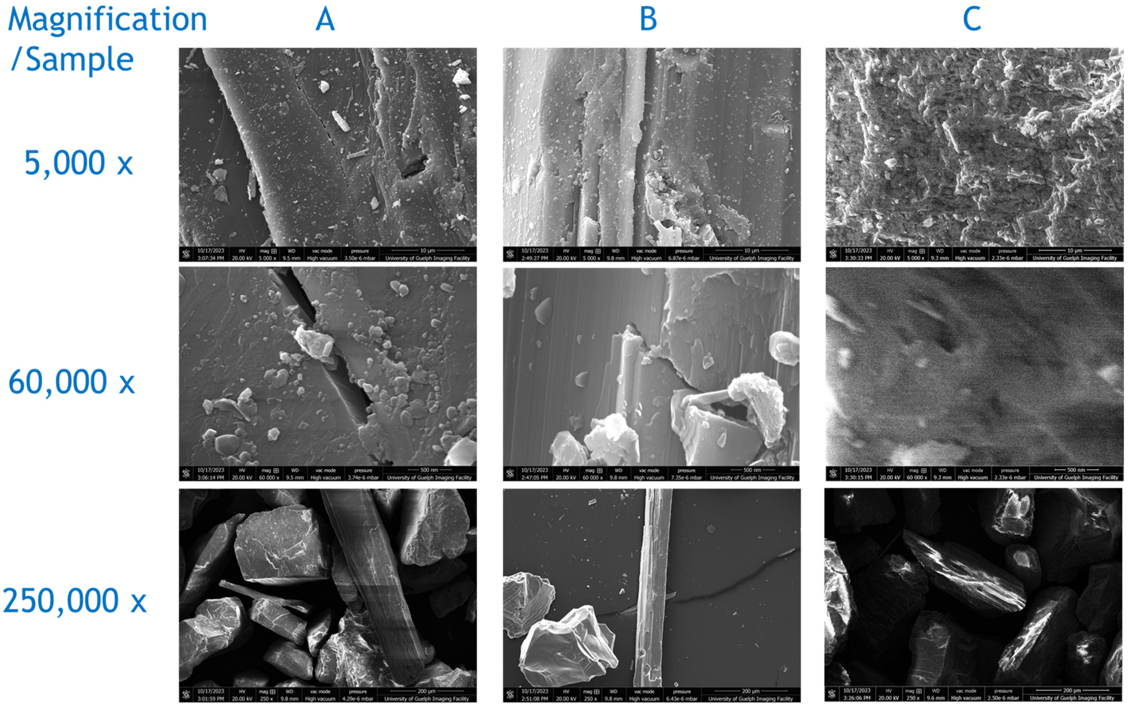
a-i Optical microscopy (first row) and FEG-ESEM (second and third rows)

By A Mystery Man Writer
Download scientific diagram | a-i Optical microscopy (first row) and FEG-ESEM (second and third rows) images of the Afghan (a, d, g), Siberian (b, e, h), and Chilean (c, f, i) lapis lazuli stones and their derived pigments (third row) from publication: Characterization of lapis lazuli and corresponding purified pigments for a provenance study of ultramarine pigments used in works of art | In this paper, we propose an analytical methodology for attributing provenance to natural lapis lazuli pigments employed in works of art, and for distinguishing whether they are of natural or synthetic origin. A multitechnique characterization of lazurite and accessory phases | Pigmentation, Paintings and Art | ResearchGate, the professional network for scientists.

Photoreceptor phagocytosis is mediated by phosphoinositide signaling - Mustafi - 2013 - The FASEB Journal - Wiley Online Library
a-i Optical microscopy (first row) and FEG-ESEM (second and third rows)

In situ ESEM using 3-D printed and adapted accessories to observe living plantlets and their interaction with enzyme and fungus - ScienceDirect

Scanning Electron Microscopy - Physics of Image Formation and Microanalysis - Ludwig Reimer (1998 Second Edition), PDF, Scanning Electron Microscope

Specialized SEM Techniques

PDF) Scanning Electron Microscopy and X-Ray Microanalysis
Applied Sciences, Free Full-Text
IJMS, Free Full-Text
Applied Sciences, Free Full-Text
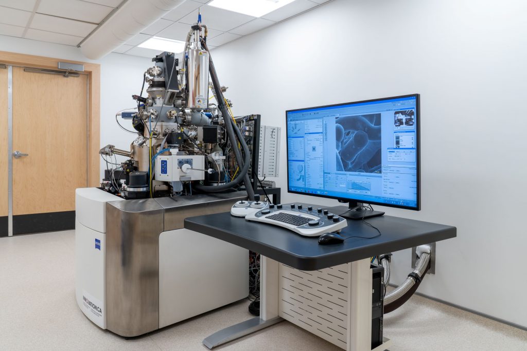
Instruments - Canadian Centre for Electron Microscopy

PDF) Characterization of lapis lazuli and corresponding purified pigments for a provenance study of ultramarine pigments used in works of art
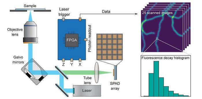
ism-microscope-1708448709720.jpg
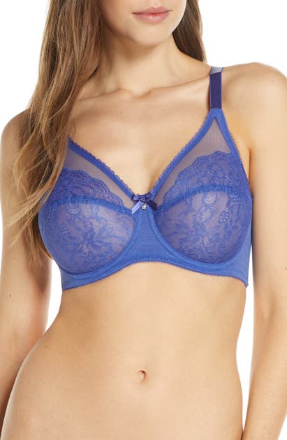
- GODDESS Verity Full Cup Underwire Bra (700204),40M,Fawn at Women's Clothing store

- Wacoal Retro Chic Full Figure Underwire Bra In Iris
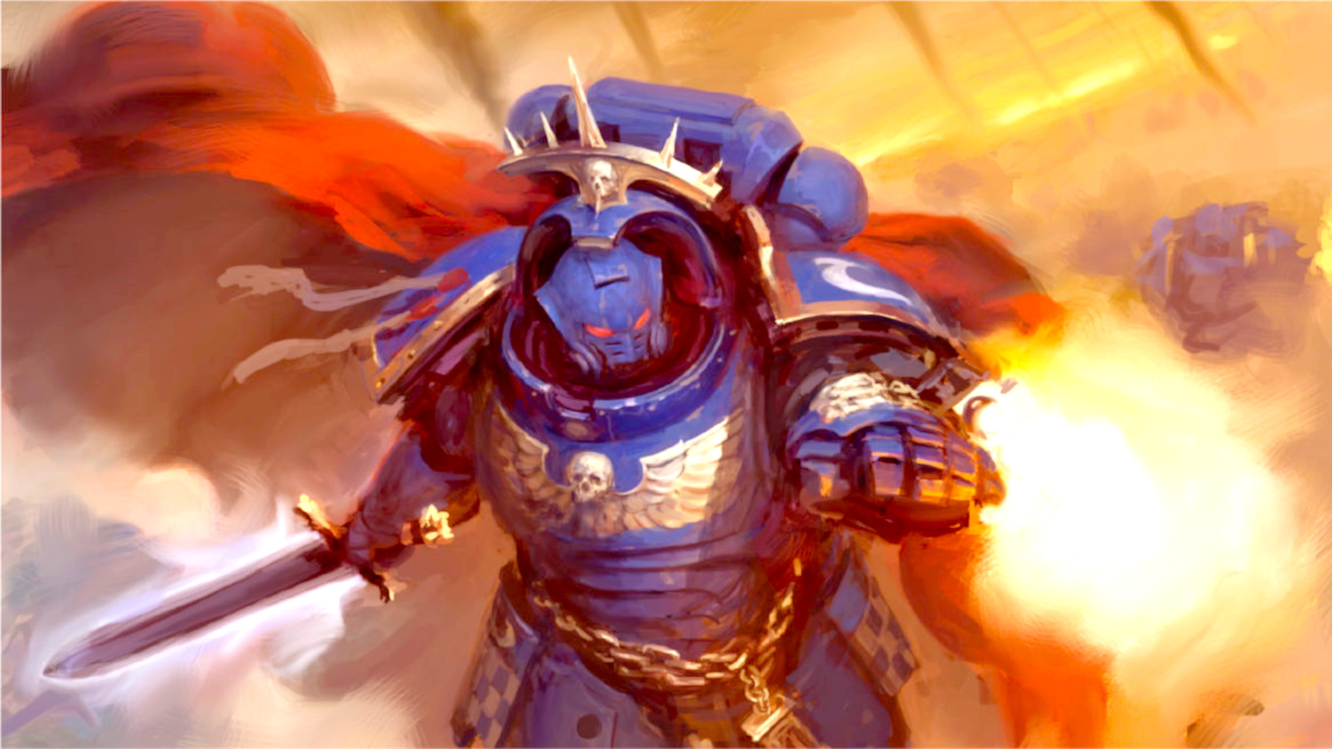
- Warhammer 40k factions – all 40k armies and races explained
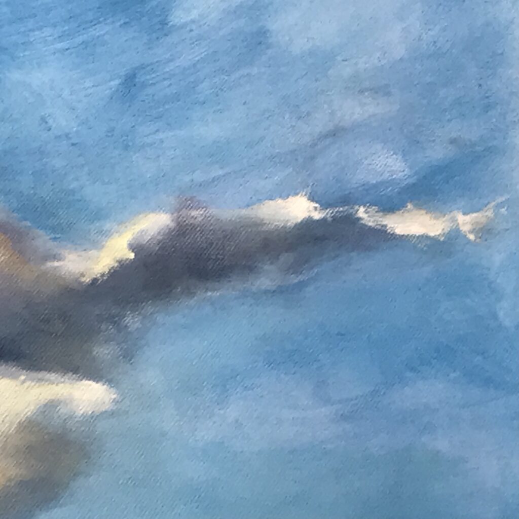
- The Story Behind the Painting – Melanie Stokes Art
- MAYBELLINE NEW YORK Color Tattoo 24H eye stix 40 I am Fierce 3v1 1,4 g - Eyeshadow

- Top View of Old Sound Recording Tape, Reel To Reel Type and Box with Room for Text. Filtered Image Stock Image - Image of disco, cover: 47476019
- Vestido Urbanic, Vestido Feminino Urbanic Nunca Usado 80089552
- Buy OOLA LINGERIE Everyday Moulded T Shirt Bra 46DD, Bras
- DORINA OUTRUN PUSH UP - Sports-bh'er - grey melange/grå

- 2-Pack Women Bra Daily Comfort (Black)