a B-mode image demonstrating a cervical length measurement. Cervical

By A Mystery Man Writer
Download scientific diagram | a B-mode image demonstrating a cervical length measurement. Cervical tissue is outlined with the dotted line. The endocervical canal is demonstrated with a solid line. Two contiguous segments are often used when the cervix is not straight. The cervical length on this patient is 37.1 mm, which is in the normal range. b Ultrasound images illustrating the assessment of cervical consistency index (CCI). The left image is without pressure applied to the cervix. The right image is with pressure applied to the cervix by the transducer. CCI = 26 mm/32.9 mm × 100 = 79%. A smaller CCI is consistent with a softer cervix. c Strain elastography makes conclusions regarding tissue stiffness through observing deformations caused by probe pressure. Each color represents the difference in compressibility relative to the adjacent area. Softer tissue appears red while firmer tissue is assigned to blue from publication: Evolving cervical imaging technologies to predict preterm birth | Preterm birth, defined as delivery at less than 37 weeks’ gestation, increases maternal-fetal morbidity and mortality and places heavy financial and emotional burdens on families and society. Although premature cervical remodeling is a major factor in many preterm deliveries, | Preterm Birth, Elasticity Imaging Techniques and Elastography | ResearchGate, the professional network for scientists.

How to measure cervical length - Kagan - 2015 - Ultrasound in

d45jl3w9libvn.cloudfront.net/jaypee/static/books/9

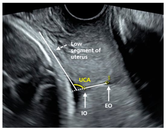
Uterocervical angle versus cervical length in the prediction of

Value of cervical strain in ultrasound elastography as a predictor
JCM, Free Full-Text

a B-mode image demonstrating a cervical length measurement

How To Measure Cervical Length On Ultrasound
Cervical Cerclage Revisited, Article

JaypeeDigital

Measurements of the cervix with funneling. (A) Funnel length, (B
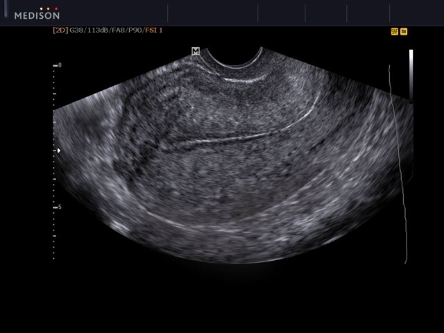
- Ultrasound images • Uterus, B-mode, echogramm №558
- FlightAware Band Pass Signal Filter, Dual 978-1090 MHz
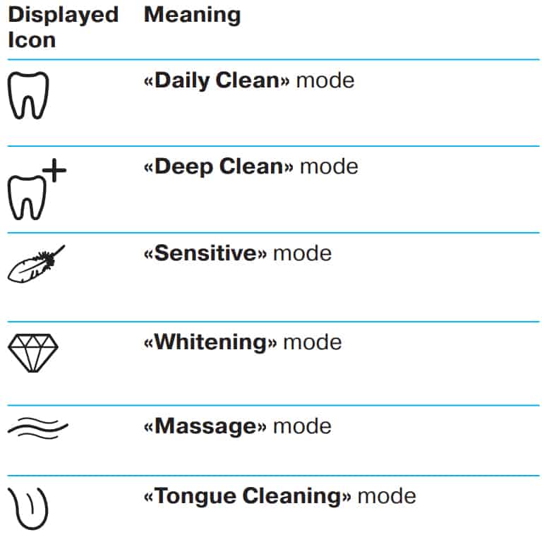
- Oral-B cleaning modes explained - Electric Teeth
- Handheld Point-of-Care Ultrasound Probes: The New Generation of POCUS - ScienceDirect

- Ultrasound imaging in B-mode, color and spectral Doppler of the
- VERSACE - STRETCH COTTON THONG WITH GREEK AND MEDUSA - Eleonora Bonucci
- Coterie Diapers, Pricing, Cost, Reviews
- Tiny waist

- Flourish_ladies_undergarments - Flourish Apple Bra l Rs.440 Color : Skin / Black / White Size : 32B / 34B / 36B / 38B / 40B / 42B / 44B Place an order via Whatsapp
- Full Support Non-Slip Convertible Bandeau Bra, Nakans Non Slip Convertible Bandeau Bra, Convertible Bandeau Bra (Black,34/75C) : : Clothing, Shoes & Accessories