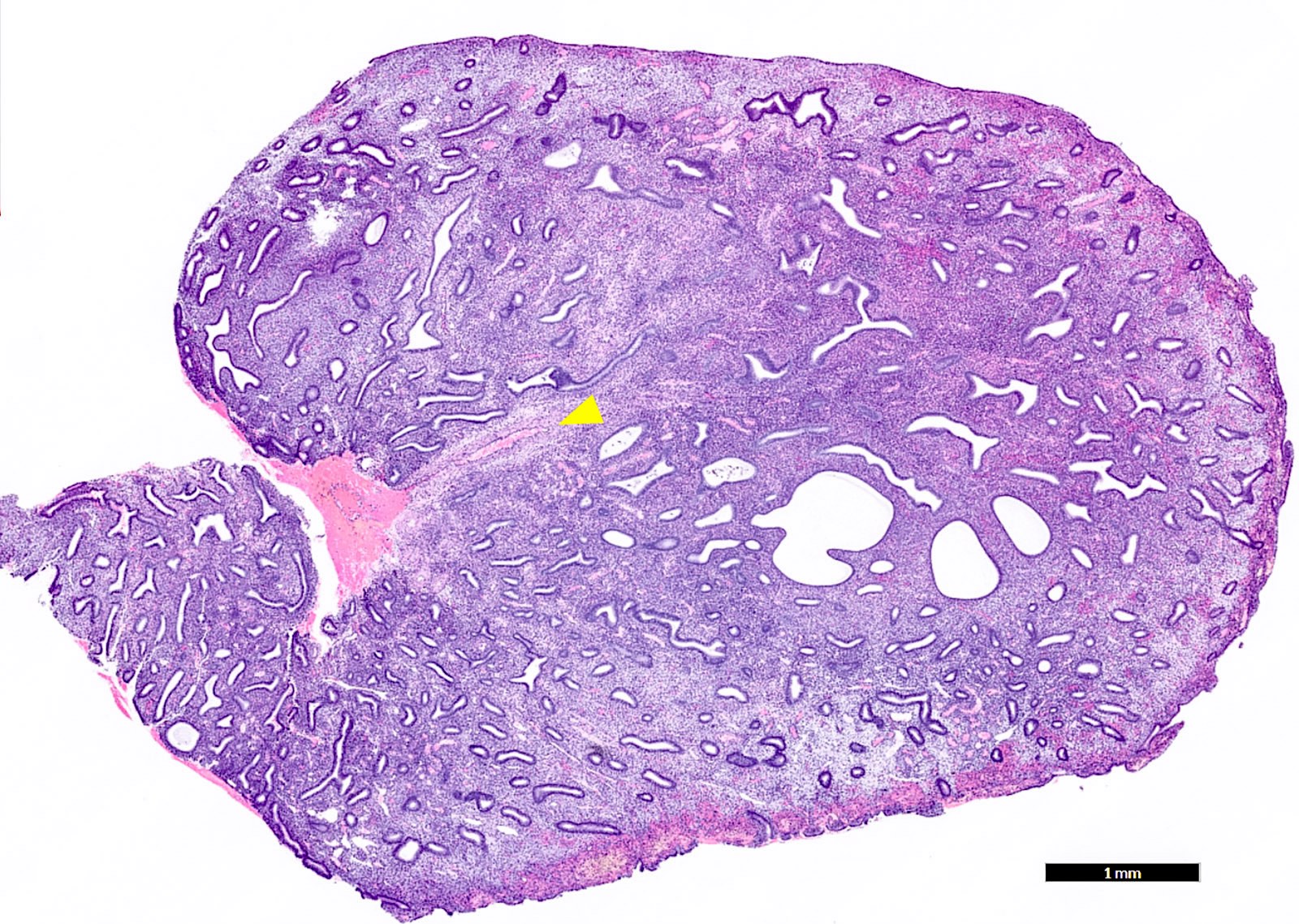
Gross image showing dilated and thick-walled blood vessels with

By A Mystery Man Writer

Markedly dilated congested blood vessels lined by a thin layer of
Histology shows a tissue composed of large, dilated, congested

Low magnification of lesion showing thick-walled myometrial

Arteriovenous Malformation of Greater Omentum: First Case Report

Vasodilation - Wikipedia
Gross image showing dilated and thick-walled blood vessels with

Photomicrograph of a pathological specimen revealing dilated

Histopathology images of pulmonary vascular disease: Thick-walled
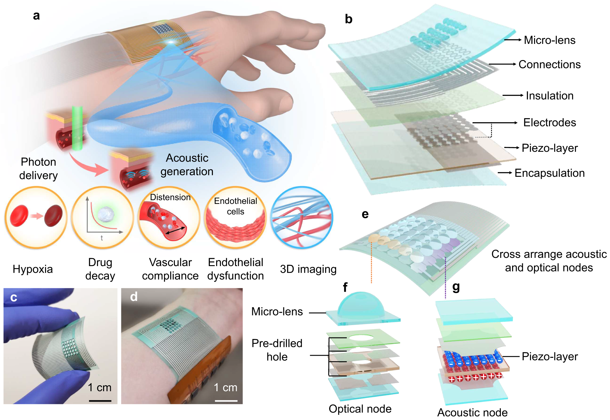
A flexible optoacoustic blood 'stethoscope' for noninvasive

Histological markers, sickle-shaped blood vessels, myxoid area
Pathology Outlines - Endometrial polyp

Vessel Diameter - an overview

Ashley STUECK Hepatobiliary & Gastrointestinal Pathologist
Mucosal edema hi-res stock photography and images - Alamy

Extracellular Matrix in Vascular Disease, Part 2/4: JACC Focus
- Sputum: Definition, colors, causes, and when to see a doctor

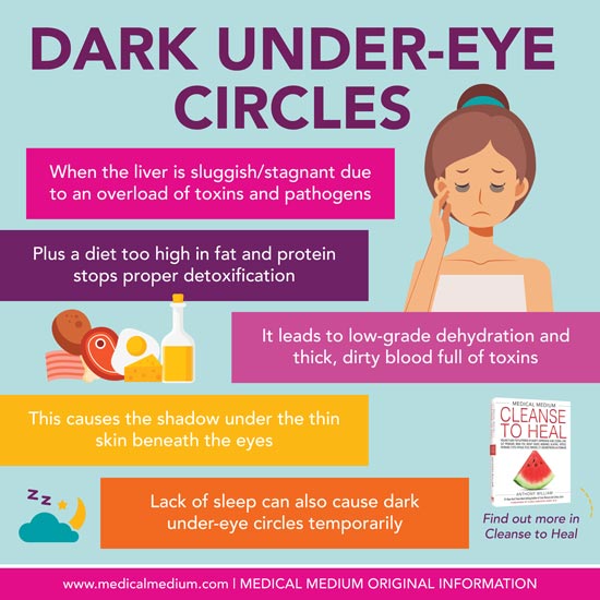
- Dark Under-Eye Circles
- DELISOUL Stage Blood 1 Fl Oz(30ml),Ultra-Realistic Fake Blood,Halloween Cosplay Special Effect Wound FX Blood for Theater,Costume Maleup,Zombie,Vampire,Monster SFX Makeup,Drips & Never Dries,Dark : Beauty & Personal Care

- Crumpled red thick paper. Bright beautiful intense blood tint. Texture. Dark vignetting around the edges of the page. Saturated color. Bulge effect Stock Photo
- BEN NYE STAGE BLOOD/ THICK/ DARK BLOOD-CHOOSE YOUR SIZE-100% GENUINE-UK SELLER