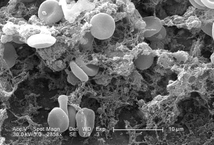
Red and white blood cells in clot, SEM - Stock Image - C045/8688 - Science Photo Library

By A Mystery Man Writer
Red blood cells (erythrocytes) and a single white blood cell (leucocyte or leukocyte) in a fibrin mesh, coloured scanning electron micrograph (SEM). Formation of a blood clot with many erythrocytes (red) and a single leukocyte (white/blue) becoming entangled in a fibrin mesh (light brown). ANNE WESTON, FRANCIS CRICK INSTITUTE/SCIENCE PHOTO LIBRARY

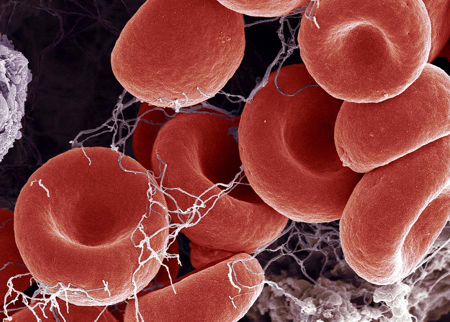
Human red and white blood cells, SEM - Stock Image - C032/0863

Red blood cells, white blood cells and platelets, SEM - Stock

Science Photo Library - Illustration of a blood clot. Depicted

Blood Clot, Sem #26 by Steve Gschmeissner

Red and white blood cells in clot, SEM - Stock Image - C045/8688
Details - Public Health Image Library(PHIL)
Blood Clot, Sem #7 by Steve Gschmeissner

Blood clot, SEM - Stock Image - F002/7300 - Science Photo Library

Red blood cells, white blood cell and platelets, SEM - Stock Image

Human red and white blood cells, SEM - Stock Image - C032/0863

Science Photo Library (@sciencephotolibrary) posted on Instagram

Human red and white blood cells, SEM - Stock Image - C032/0863

Blood Clot, Sem #26 by Steve Gschmeissner
- Under Armour Unveils Curry Two 'Red, White & Blue' [PHOTOS]

- Vintage White Ironstone Soup Tureen with Ladle and Under Plate, Grape - Ruby Lane

- Tênis Under Armour UA Charger Slight Se Psh Pink/Red Fus/White
- Grunge Red Under Review Word Round Rubber Seal Stamp On White

- I still have no idea why red is best under white, but here's the proof that it works! 😍 There are some beautiful red bra options for you…