Ultra-wide-field fundus photographs and ultra-wide-field

By A Mystery Man Writer
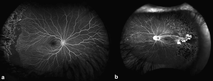
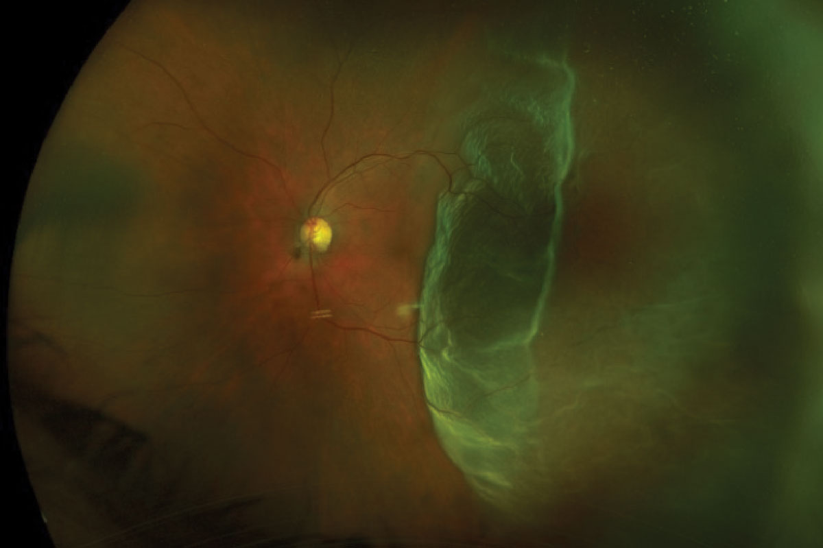
Download scientific diagram | Ultra-wide-field fundus photographs and ultra-wide-field fluorescein angiographic imaging of ocular toxocariasis. (A) A granuloma with mild vitreous opacity. (B) A tractional retinal fold with localized tractional retinal detachment. (C) Diffuse peripheral vascular leakage. (D) A prominent optic disc leakage. from publication: The Clinical Characteristics of Ocular Toxocariasis in Jeju Island Using Ultra-wide-field Fundus Photography | Toxocariasis, Ocular and Photography | ResearchGate, the professional network for scientists.

Comparison of true-colour wide-field confocal scanner imaging with standard fundus photography for diabetic retinopathy screening
The utility of ultra-widefield fluorescein angiography in pediatric retinal diseases, International Journal of Retina and Vitreous
PDF) The Clinical Characteristics of Ocular Toxocariasis in Jeju Island Using Ultra-wide-field Fundus Photography

ZEISS CLARUS 500 Fundus Camera

Eun Kyoung Lee's research works Dongguk University, Seoul and other places
Ultra-Wide Field Retinal Imaging Device, Product Technology
DrushtiEye and Retina Center:-Usefullness Or Ultrawide Field Fundus Photo With Periferal Swept Source Oct

Figure 2 from Emerging Issues for Ultra-Wide Field Angiography.
Ultra-Widefield Imaging: Expand Your Horizons


- Women's Ladies Formal Office Trouser or Workwear Pant Ash Blue

- Floerns Women's Off Shoulder Long Sleeve Lace Up Back Bandeau

- atomicdogz77 on X: I'll give you jeans and a hint of pantyhose

- KUAK Yoga Mat Bag Large Yoga Bags and Carriers with Yoga Mat Strap, Full Zipper Closure, 5 Multi-Functional Pockets, Durable Yoga Mat Tote Sling Carrier for Women Fits Most Size

- Cotton Wire-Free Bra - 2 Pack